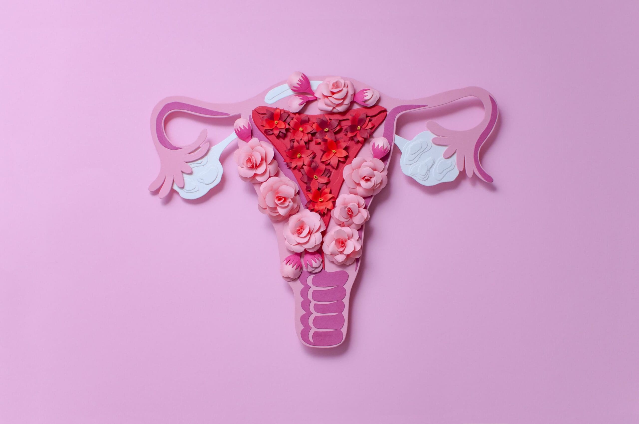
The female reproductive organs include the ovaries, fallopian tubes, uterus and vagina.
Front view of female reproductive organs

Side view of female reproductive organs

Ovaries
The ovaries are the main reproductive organs of a woman. The two ovaries, which are about the size and shape of almonds, produce female hormones (oestrogens and progesterone) and eggs (ova). All the other female reproductive organs are there to transport, nurture and otherwise meet the needs of the egg or developing fetus.
The ovaries are held in place by various ligaments which anchor them to the uterus and the pelvis. The ovary contains ovarian follicles, in which eggs develop. Once a follicle is mature, it ruptures and the developing egg is ejected from the ovary into the fallopian tubes. This is called ovulation. Ovulation occurs in the middle of the menstrual cycle and usually takes place every 28 days or so in a mature female. It takes place from either the right or left ovary at random.
Fallopian tubes
The fallopian tubes are about 10 cm long and begin as funnel-shaped passages next to the ovary. They have a number of finger-like projections known as fimbriae on the end near the ovary. When an egg is released by the ovary it is ‘caught’ by one of the fimbriae and transported along the fallopian tube to the uterus. The egg is moved along the fallopian tube by the wafting action of cilia — hairy projections on the surfaces of cells at the entrance of the fallopian tube — and the contractions made by the tube. It takes the egg about 5 days to reach the uterus and it is on this journey down the fallopian tube that fertilisation may occur if a sperm penetrates and fuses with the egg. The egg, however, is usually viable for only 24 hours after ovulation, so fertilisation usually occurs in the top one-third of the fallopian tube.
Uterus
The uterus is a hollow cavity about the size of a pear (in women who have never been pregnant) that exists to house a developing fertilised egg. The main part of the uterus (which sits in the pelvic cavity) is called the body of the uterus, while the rounded region above the entrance of the fallopian tubes is the fundus and its narrow outlet, which protrudes into the vagina, is the cervix.
The thick wall of the uterus is composed of 3 layers. The inner layer is known as the endometrium. If an egg has been fertilised it will burrow into the endometrium, where it will stay for the rest of its growth. The uterus will expand during a pregnancy to make room for the growing fetus. A part of the wall of the fertilised egg, which has burrowed into the endometrium, develops into the placenta. If an egg has not been fertilised, the endometrial lining is shed at the end of each menstrual cycle.
The myometrium is the large middle layer of the uterus, which is made up of interlocking groups of muscle. It plays an important role during the birth of a baby, contracting rhythmically to move the baby out of the body via the birth canal (vagina).
Vagina
The vagina is a fibromuscular tube that extends from the cervix to the vestibule of the vulva. The vagina receives the penis and semen during sexual intercourse and also provides a passageway for menstrual blood flow to leave the body.


