
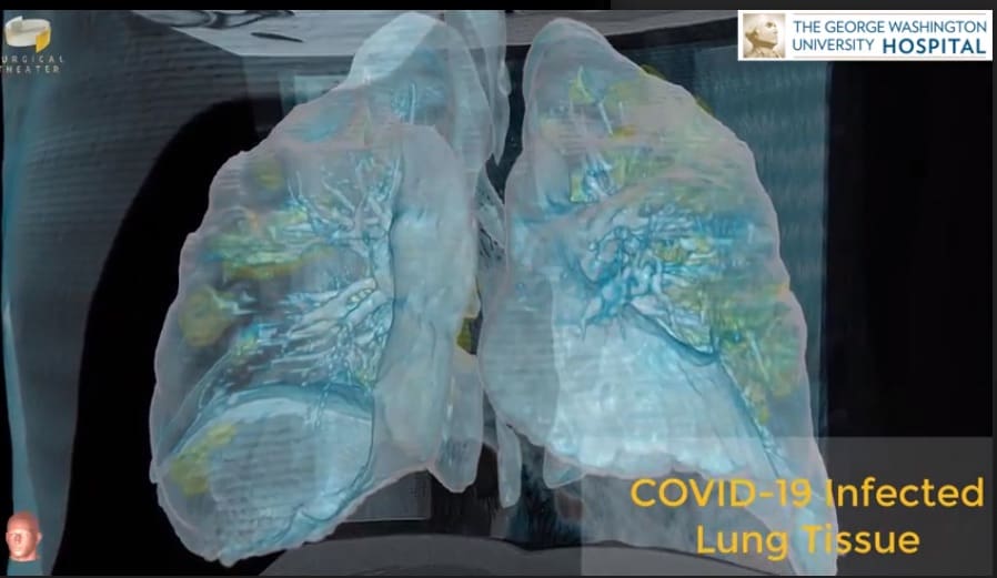
Click the image to see inside the lungs of a patient with COVID-19 in this virtual reality 3-D video created by George Washington University Hospital using Surgical Theater technology.
Damage to the lungs is shown in yellow.
The patient was a man in his late 50s transferred from another hospital when his initial symptoms (fever, cough, shortness of breath) quickly escalated. He was connected to a ventilator, but then required ECMO – extracorporeal membrane oxygenation – a technology which removes blood from the body, oxygenates it and then returns it to the body.
“There is such a stark contrast between the virus-infected abnormal lung and the more healthy, adjacent lung tissue,” said Dr Keith Mortman, Chief of Thoracic Surgery at George Washington Hospital.
“And it’s such a contrast that you do not need an MD after your name to understand these images. This is something the general public can take a look at and really start to comprehend how severe the amount of damage this is causing the lung tissue. The damage we’re seeing is not isolated to any one part of the lung. This is severe damage to both lungs diffusely.”
Dr. Mortman is especially concerned with the possibility of enduring damage to the lungs of those who survive COVID-19. “When that inflammation does not subside with time, then it becomes essentially scarring in the lungs, creating long-term damage,” he said. “It could impact somebody’s ability to breathe in the long term.”


